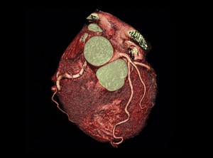
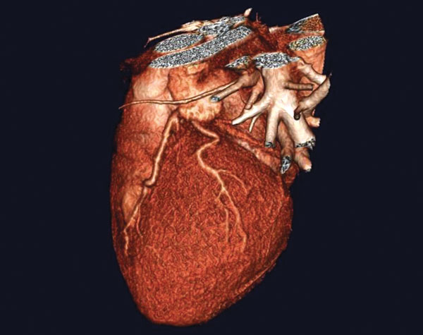
CT Coronary Angiogram identifies blockages inside the artery (hard plaque) and inside the artery wall (soft plaque)
Advances in technology over the past 30 years have allowed clinicians to diagnose and treat patients using less invasive and more accurate methods. The introduction of Magnetic Resonance Imaging (MRI), Computed Tomography (CT) and Positron Emission Tomography (PET) changed the way physicians “looked inside” a body to see if there was an abnormality. Over the years, technologically-advanced imaging has evolved to detect disease in a minimally invasive manner and with greater accuracy than traditional screening methods.
Over the past 5 years, the technology has developed even further. Today, MRI, CT and PET/CT can now effectively screen for the following diseases:
Coronary Artery Disease
Coronary artery disease is the leading cause of death for men and women worldwide. Historically, physicians would perform a coronary catheter angiogram to check for blockage of the arteries. This method, while effective, requires that a patient be sedated and have a small incision made in their groin for the catheter. Today, the same exam can be performed with CT and doesn’t require sedation or a catheter. Unlike a traditional angiogram which only sees blockage inside the artery, the CT angiogram identifies blockage inside the artery (hard plaque) and inside the artery wall (soft plaque). Soft plaque is the “silent killer” as it is hidden inside the artery wall and can cause sudden death if it ruptures. In 30% of patients who have coronary artery disease, their first symptom is sudden death.
Prostate Cancer
Prostate cancer is the second most common cancer in American men, and 1 in 6 will be diagnosed with prostate cancer in their lifetime. Currently, the PSA (prostate-specific antigen) blood test is the most widely used screening tool for prostate cancer. While the PSA is useful in detecting a problem in the prostate gland, it is not specific enough to determine if it is elevated due to benign disease or cancer. Historically, urologists would perform a trans-rectal ultrasound guided biopsy (TRUS) to identify the cause. Unfortunately, because TRUS biopsies are done by randomly sampling the prostate gland, they do not detect lesions 35% of the time because they can’t see the tumor inside the gland. With the advances in MRI technology, radiologists and physicians can now see inside the gland and identify tumor suspicious areas with pinpoint precision. This new exam is called a multiparametric MRI of the prostate. This same technology is also used to guide biopsy of a concerning lesion to determine if it is cancer. If cancer is found early, a patient may be able to have focal treatment of the cancer versus whole gland therapy.
Colon Cancer
Colon cancer is the third most common type of cancer, and it has a very high survival rate if caught early. The current recommended guideline for screening is to get a colonoscopy beginning at age 50. Traditional colonoscopies are performed by inserting a thin flexible scope into the colon to look for polyps or tumors. Patients are sedated for their comfort. A less invasive alternative is a Virtual Colonoscopy performed with CT and 3D “fly through” reformations. This exam requires no sedation and identifies polyps or masses inside of the colon, while also producing images of surrounding abdominal and pelvic organs, allowing for their interrogation as well.
Alzheimer ‘s Disease
Alzheimer’s disease affects 1 in 8 Americans over the age of 65. While there is currently no cure for Alzheimer’s, early detection can help patients and their families plan for their future. Researchers have found that a diagnosis of Alzheimer’s is supported when there is atrophy of the hippocampal formations. Today, there is a diagnostic MRI with 3D volumetric quantitative analysis (NeuroQuant) that can measure the volume of the hippocampus, temporal and parietal lobes and other structures of the brain. Most recently, the FDA approved an isotope called Amyvid for PET/CT that specifically identifies amyoid plaques in the brain which are associated with Alzheimer’s disease. These plaques are thought to hinder normal brain function and promote degeneration of brain tissue.
Advanced medical imaging is a great solution for patients who may choose not to undergo standard screening tests due to anxiety related to invasive procedures. Most physicians agree that the best test a patient can have is the one they are willing to undergo.
Dr. Hancock is a Board-Certified Neuroradiologist at Desert Medical Imaging. DMI performs all these screenings at their Indian Wells facility and can be reached at 760.694.9559.


Comments (0)