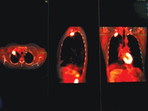
PET/CT demonstrating a lung cancer in the right lung apex and normal heart uptake
PET/CT is an acronym that stands for “positron emission tomography/computed tomography.” It is a combination of PET and CT (CAT scan) which combines the best of both worlds in terms of functional metabolic imaging at the cellular level, and anatomic imaging for structural detail. It also incorporates the PET scan’s ability to use special probes (also called agents or tracers) to identify areas of abnormality.
In cancer imaging, PET/CT can be useful to map out locations of old or new cancer growth. Cancer cells reproduce rapidly and in order to do that, they consume glucose (a form of sugar). The faster the cancer cells grow, the more glucose they consume. FDG is a safe and effective form of glucose that emits particles called positrons; when FDG is injected into a patient, it becomes concentrated in areas of rapid consumption. A PET/CT scan detects these areas of increased FDG and maps the abnormal area onto a CT image showing structural detail. This helps the physician detect the presence of disease and localize it precisely.
For these reasons, PET/CT is commonly used in cancer imaging; it is also used in cardiology to image the part of a heart that may have suffered damage following a heart attack. PET/CT can also demonstrate coronary artery disease before the onset of symptoms, and show if a cardiac treatment has been effective. PET/CT can also be useful in better understanding brain metabolism, particularly for Alzheimer’s disease, epilepsy and Parkinson’s.
A PET/CT scan is a simple process which can significantly help your health care team identify issues and guide your treatment and care plan. The scan process includes some preparation such as no eating or drinking for 6 hours before your study, and no heavy exercise for one day prior to your exam. At the time of the examination, an I.V. will be placed and a painless injection will be administered. Patients lie quietly (without speaking) for 60 minutes allowing the FDG to distribute in the body. After the scan is completed, you may leave and resume normal activity. A report will be sent to your doctor by an expert radiologist, and your doctor will contact you with the results.
Bernadette Greenwood is director of clinical services at Desert Medical Imaging (DMI), as well as an author and educator. Dr. Brochert is a board-certified radiologist at DMI who specializes in PET and cancer imaging. For more information visit www.DesertMedicalImaging.com or call
(760) 694.9559.


Comments (0)