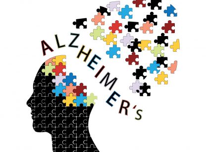
Alzheimer’s disease affects one in eight Americans over the age of 65. Approximately 5.4 million Americans suffer from Alzheimer’s disease and 82 million are expected worldwide by 2050.
Unfortunately there is still no cure for Alzheimer’s but researchers are continually working to understand the hallmarks of the disease so that they can find a cure. One of the most difficult aspects of their studies is that most of their results are determined through pathology after the patient has died. A more effective way to study the cause and effect of different types of therapies is to measure the results “in vivo,” while the patient is alive.
In recent years Advanced Imaging has become an essential tool for identifying areas of the brain that are affected when dementia is present. These tools assist physicians in diagnosing Mild Cognitive Impairment (MCI) from Alzheimer’s disease and other dementia causing etiologies and are performed while the patient is living. Currently, there are three different types of imaging that are showing great promise in diagnosing Alzheimer’s.
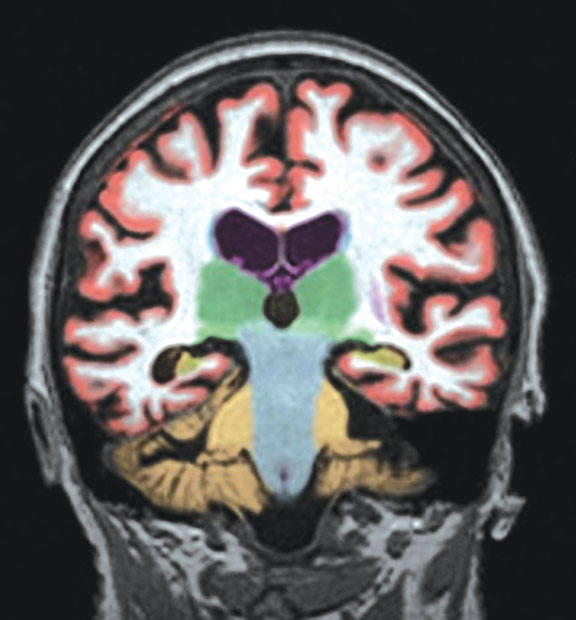
Structural Imaging looks at the brain as a whole and looks for changes in shape, position or volume of the brain. Today, there is a diagnostic MRI with 3D volumetric quantitative analysis (NeuroQuant) that can measure the volume of the hippocampus, temporal and parietal lobes and other structures of the brain. Alzheimer’s occurs when there is atrophy (shrinkage) of the volume of the brain, and specifically the hippocampus. Structural imaging can also be used to help discern Alzheimer’s from other potential entities such as vascular dementia, Huntington’s disease, Parkinson’s disease and pseudo-dementia related to major depression.
Functional Imaging looks at how well the cells in various brain regions are working by showing how actively they are using sugar or oxygen. PET/CT is used with fluorodeoxyglucose (FDG) to find areas of the brain that have reduced use of sugar which is often associated with memory, learning and problem solving.
Molecular Imaging uses highly targeted radiotracers to detect cellular or chemical changes linked to a specific disease. Most recently, the FDA approved an isotope called Amyvid for PET/CT that specifically identifies amyloid plaques in the brain which are associated with Alzheimer’s disease. These plaques are thought to hinder normal brain function and promote degeneration of brain tissue. While the presence of plaque isn’t a definitive diagnosis of Alzheimer’s as a small amount of plaque is common in older people, the absence of plaque is confirmation that the patient does not have Alzheimer’s and that their mental decline is due to another cause.
These exams may be used clinically to diagnosis a patient and by researchers to determine the effectiveness of new drugs in hopes of finding a cure.
As our Baby-boomers continue to age, Alzheimer’s will become a more common disease. However, just like other diseases, Alzheimer’s can be alleviated by managing your physical health. You can reduce the risk of cognitive impairment with physical activity, a healthy diet, keeping your mind active, maintaining healthy blood sugars and good heart health, while reducing salt intake and alcohol consumption.
Dr. Hancock is a Board Certified Neuroradiologist and can be reached at Desert Medical Imaging (760) 694.9559. www.desertmedicalimaging.com


Comments (0)