I always fill out the forms quickly in the doctor’s waiting room. Do you have a history— No. Have you ever— No. I didn’t have a family history of breast cancer or any cancer. But a year ago this past December, my mother was diagnosed and ended up receiving a double mastectomy. The key is to catch it early. And really the only way you can catch breast cancer early is through screening technology.
Before my mom’s diagnosis, I would occasionally get mammograms. But that was before. To understand mammography and recent developments in screening, I met with Dr. Marla Lander, a fellowship-trained radiologist and breast imager at the Comprehensive Cancer Center at Desert Regional Medical Center. The first question: is the radiation used dangerous?
“No,” she said. The dose a woman gets during a screening mammogram with two views is about the same from the environment over about 7 weeks. Then we got to the hard questions: why is there so much negative press around mammography?
“It does make a huge difference, but it’s not perfect. It can miss cancers in women with dense breast tissue,” Dr. Lander said. Only about 40 percent of breast cancers present with calcifications. “You’ll catch these with mammography, but that leaves the other 60 percent that can be tricky to find on the mammogram if the woman has dense breast tissue.”
The problem with dense breasts is that it makes detecting cancer more challenging because cancer can be hidden in the tissue. After menopause or after a woman stops hormone replacement therapy, dense breasts will typically transition to fatty breasts. However, for 20 percent of women, dense breast tissue will remain an issue throughout their lives. The major risk factor, then, is missing detection of these hidden cancers during a mammogram.
How do you detect cancers in dense breast tissue? With 3-D imagery. “Screening ultrasound is revolutionizing how we practice breast medicine,” said Dr. Lander. The Comprehensive Cancer Center has a Somo-v automated 3-D breast ultrasound that can acquire 3-D images of dense breasts in less than 5 minutes.
“The automated system covers every inch of the breast,” said Dr. Lander. “And it’s reproducible. You can compare the results side by side and from one year to the next. That’s a huge advantage.”
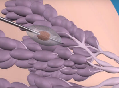
For example, there is one particular type of breast cancer that accounts for about 15 percent of cases. “It’s easy to miss on a mammogram, MRI or even a PET scan,” said Dr. Lander. “A fantastic advantage of the coronal view ultrasound is that lobular cancers jump out at the reader.”
For a patient like myself, this was all I needed to know. I scheduled both a mammogram and a 3-D ultrasound screening at the Comprehensive Cancer Center. I got passing marks and peace of mind.
And I called my mom.
For more information about the Comprehensive Cancer Center, call (760) 416.4800 or visit www.desertcancercenter.com/breasthealth.


Comments (0)